02
2025/12/17 Minerva Technology – 3D SEM Reconstruction and SYM Cuesta Section Collection System Workshop
2025.11.10
Introduction:
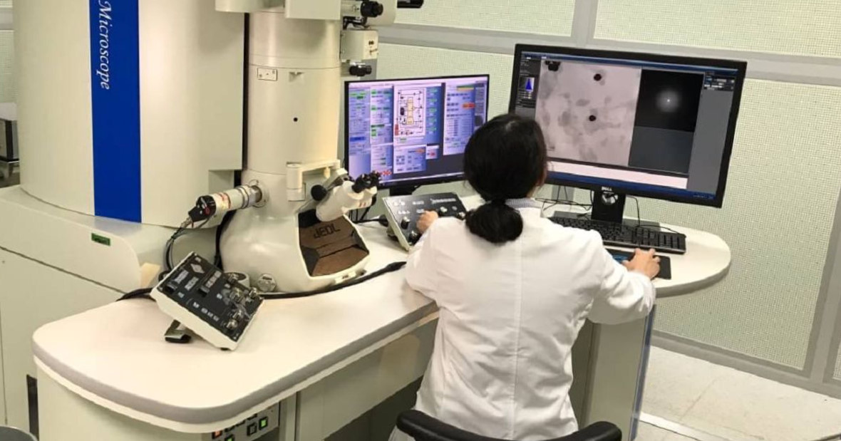
After
decades of advancement, the electron microscope has become an indispensable
tool in modern scientific research. Ultrathin sectioning technology is an
essential step in electron microscopy sample preparation. In recent years, with
technological progress, ultrathin sectioning has evolved—from room temperature
to cryogenic conditions, and from two-dimensional to three-dimensional imaging.
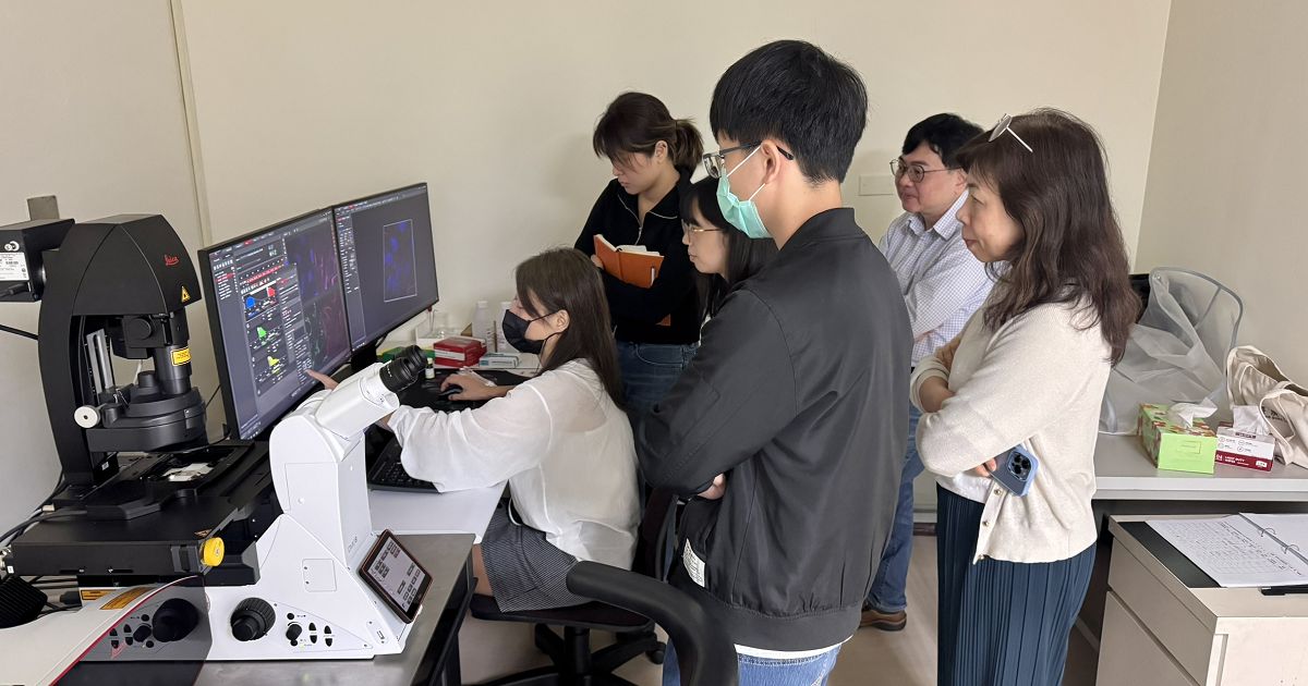
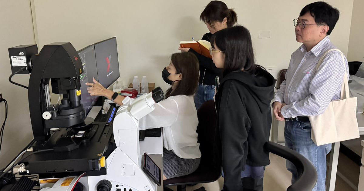
This
workshop focuses on introducing the complete workflow of SEM 3D ultrathin
section reconstruction, including sample preparation, section collection, and
SEM imaging reconstruction.
We welcome all faculty members and students to attend this section collection system
workshop. If you have any questions, please contact Ms. He at extension #5065.
=========================================
【Workshop Schedule】:
◆3D
Sample Preparation and SYM Cuesta Section Collection System (Oral Presentation)
:
Date: Wednesday,
December 17, 2025
Time: 10:00 AM –
11:00 PM
Venue: 4th Floor,
Area B, Room 0459, Microscopy Center, First Medical Building, Chang Gung
University
Speaker: Mr. Shao-Hsuan Lin,
Instrument Specialist, Minerva Technology
◆Next-Generation
Biological Scanning Electron Microscopy: Volume EM and Correlative Microscopy
(Oral Presentation):
Date: Wednesday, December 17, 2025
Time: 11:00 AM –
12:00 PM
Venue: 4th Floor, Area B, Room
0459, Microscopy Center, First Medical
Speaker: Mr. Kang-You Fan, Application Engineer, Carl Zeiss
◆Hands-on Practice:
Date: Wednesday,
December 17, 2025
Time: 14:00 PM –
17:00 PM
Venue: 4th Floor,
Area B, Room 0459, Microscopy Center, First Medical Building, Chang Gung
University
Speaker: Mr. Shao-Hsuan Lin,
Instrument Specialist, Minerva Technology
※Notice
Due
to limited space, early registration is recommended.It is suggested that each
laboratory send at least one representative to participate.
Registration
Link:
https://forms.gle/iLweuu9SZ28uz6zA8
※Kindly respond before December 16 (Tuesday).
Thank you for your cooperation.


![[Instrument operation video] Stellaris8 basic operation teaching video](/Uploads/upload/9db7f1bd-3b3f-4d81-90d0-e7f4e38969e4.jpg)
